Uterine Malformations and Pregnancy
Congenital uterine malformations can occur as a result of the fusion and/or resorption of Muller ducts during organogenesis. Their incidence is 3-5%. The consequences of these anomalies on gynaecological and especially obstetrical pathology make the study of embryogenesis and organogenesis all the more important. Also, timely detection is a very good reason why a woman’s reproductive organs should be examined (para)clinically from as early as her birth.
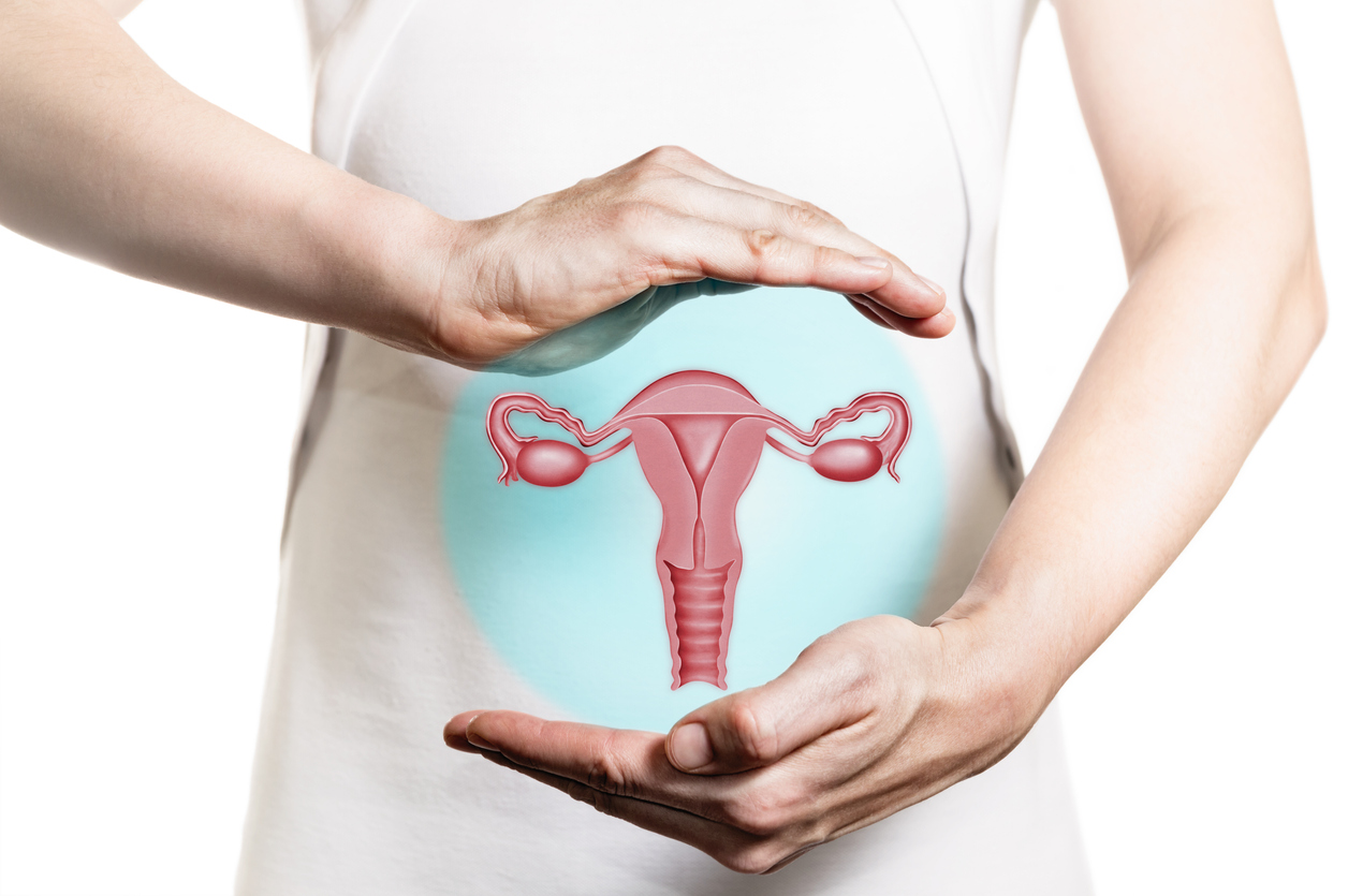
The uterus is a hollow organ shaped as an upside down pear 6-8 centimetres tall and 3-4 centimetres wide and which, by the end of a pregnancy, can reach 30 centimetres in diameter. The lower part of the uterus connects with the vagina as is called a cervix. Located on each side of the uterine fundus, the fallopian tubes direct the oocytes and the spermatozoa to meet and achieve fertilisation.
When the uterus differs in shape and structure from as early as the woman’s birth, we call that a congenital uterine malformation. Uterine anomalies may also occur later in life as a result of infections or treatments. Most women with uterine anomalies are unaware that they have them and only discover this incidentally during routine visits to the gynaecologist or in the context of detailed investigations in case of infertility.
The most common complications of congenital uterine malformations are: infertility, spontaneous abortion, premature delivery or labour dystocia (in case of full term pregnancies). For instance, the risk of premature delivery in a patient with septate uterus is twice as high.
That is why it is important to diagnose any uterine malformations as early, completely and accurately as possible and to correct them surgically before pursuing pregnancy. At present, the most frequently used diagnostic methods are three-dimensional ultrasound, hysterosalpingography, magnetic resonance imaging (MRI), diagnostic hysteroscopy (uterine endoscopy).
The most common uterine malformations are:
1.The septate uterus characterised by internal compartmentalisation by a muscular or fibrous wall called septum, amounts to half of all cases. The septate uterus may lead to foetal malformations. In the first trimester of pregnancy, the risk of spontaneous abortion in patients with septate uterus is estimated at 28-48%, and then the risk decreases to 5% during the second trimester. The diagnosis can be easily made by means of a 3D ultrasound scan and the treatment consists in sectioning the septum with the help of a hysteroscope.
2.The unicornuate uterus is half the size of a normal uterus and only one fallopian tube. A pregnancy in such a case is possible, but this type of uterus creates many risks of complications during the pregnancy, such as preterm delivery.
3.The bicornuate uterusis rare and shaped like a heart, with a separation in the upper part giving the impression of the two horns. Women with a bicornuate uterus often suffer spontaneous abortions, but 60% of women with this malformation can give birth to a live baby.
4.The didelphic uterus is a double uterus with two vaginas separated by a septal wall and each fallopian tube attached to the ipsilateral uterus. The didelphic uterus is compatible with pregnancy.
5.The arcuate uterus does not pose any problems and is, in fact, a normal uterus except for an indentation in the upper part, which does not prevent the woman to become pregnant or to carry a pregnancy to term.
References:
Acién P. (1997) Incidence of Müllerian defects in fertile and infertile women. Hum Reprod 12(7), pp. 1372-1376.
Cfian M. (2011) Studiu clinic și statistic al evoluției sarcinei pe uterul malformat. Jurnalul de Chirurgie Iaşi, Vol. 7(2), pp. 232-237.
Managementul gravidelor cu malformaţii uterine congenitale – prezentare de cazuri (available online at http://revistaginecologia.ro/system/revista/1/38-42.pdf)
Kurjak A., Chervenak F.A., Vlădăreanu R. (2012) Tratat de ultrasonografie în Obstetrică Ginecologie, Donald School, 3rd edition, Amaltea Medical Publishing House, Bucureşti, pp.746-806.
Dr Roxana Diaconu, junior doctor, Obstetrics-Gynaecology